

What does wrist arthroscopy involve?
Wrist arthroscopy is a surgical technique that allows the inside of the joint to be seen through incisions of a few millimetres. A very fine camera, the arthroscope, is introduced through one of them, projecting a magnified image onto a screen. Small instruments are introduced through the others, used to examine and, if appropriate, treat the injury. This gives access to the cartilage, the carpal ligaments and the triangular fibrocartilage without needing to open the joint.
In the wrist, arthroscopy has two uses. As a diagnostic tool, it allows direct examination of structures that MRI does not always show clearly, and assessment of whether a ligament is stable or whether the cartilage is damaged. As a therapeutic tool, it allows certain injuries to be treated with less disruption to the tissues surrounding the joint. It is one of the standard techniques in wrist surgery and is frequently combined with other procedures.
When is it indicated?
Arthroscopy is considered when wrist pain persists despite conservative treatment and examination and imaging are not enough to decide on treatment, or when the injury can be treated better by this route. The most frequent indications are:
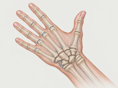
- triangular fibrocartilage injuries with pain on the little-finger side of the wrist;
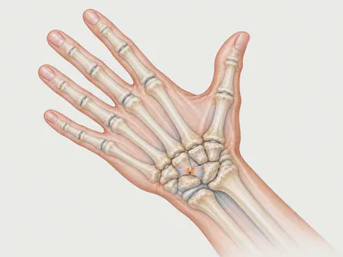
- wrist ligament injuries, particularly of the scapholunate ligament, to grade the instability and repair it when possible;
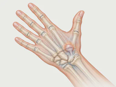
- dorsal ganglion cysts, in selected cases;
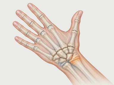
- assistance in fractures of the distal radius and scaphoid, to check that the joint surface is well reduced and to detect associated ligament injuries;
- assessment of the cartilage in early wrist osteoarthritis before deciding on another type of surgery;
- joint washout, removal of loose fragments and synovectomy in inflammatory conditions.
It does not replace examination or imaging: it is indicated when it provides information or treatment that these do not offer.
Before the procedure
The decision is based on the clinical history and examination of the wrist, with specific manoeuvres for each ligament and for the triangular fibrocartilage. X-rays are reviewed and, depending on the case, MRI or MR arthrography. In fractures, a CT scan is often useful to plan the reduction.
At the consultation, what is expected to be found, what steps might be carried out during the same procedure and what immobilisation you will need afterwards are explained, because much of the recovery depends on this. Pre-operative assessment and the anaesthesia visit are organised as for any planned surgery. If you take anticoagulants or antiplatelet medication, it is worth mentioning this in advance.
How is it carried out?
The procedure is almost always carried out as day-case surgery, with regional anaesthesia of the arm and sedation. The arm is placed in a traction tower that holds the fingers and slightly opens the joint space to allow work inside the wrist.
The access points are called portals. These are incisions of a few millimetres on the back of the wrist, between the extensor tendons, named according to their position relative to the extensor compartments. Radiocarpal portals are used for the joint between the radius and the first row of carpal bones, and midcarpal portals for the joint between the two rows of carpal bones. Fluid is introduced through them to distend the joint and improve the view.
The examination is systematic: cartilage of the radius and carpal bones, the scapholunate and lunotriquetral ligaments, the triangular fibrocartilage and the synovial membrane. Depending on the finding, the planned step is carried out: smoothing or repairing the triangular fibrocartilage, repairing or reinforcing a ligament, removing a cyst from the inside, removing inflamed tissue or checking the reduction of a fracture while it is fixed with wires or screws. At the end, the portals are closed with a stitch or an adhesive strip and a bandage or splint is applied.
Recovery and follow-up
Recovery depends mainly on what was done inside the wrist, not on the incisions, which heal quickly.
- After a diagnostic arthroscopy, a joint washout or removal of a cyst, a bandage is worn for a few days and mobilisation of the fingers and wrist starts soon.
- After repair of the triangular fibrocartilage or a ligament repair, immobilisation with a splint or cast is needed for several weeks to protect the repair, followed by rehabilitation with a hand therapist.
- After a fracture assisted by arthroscopy, the standard protocol for fracture surgery is followed.
Follow-up appointments allow the stitches to be removed, the immobilisation to be adjusted and the pace of return to work and sport to be set, which varies considerably depending on the activity and the procedure carried out.
Risks and limitations
Arthroscopy is a safe technique in trained hands, but like any surgery it carries risks: infection, irritation or injury to the sensory nerve branches or tendons passing near the portals, stiffness, persistent pain or temporary accumulation of fluid in the tissues.
It is worth being clear about its limitations. Not all injuries can be treated by arthroscopy: chronic ligament instabilities or extensive injuries may require open surgery, and this is sometimes decided during the procedure itself. Lost cartilage does not recover, so in advanced osteoarthritis arthroscopy offers little benefit. And in some cases the examination does not find a treatable injury, which is also useful information for guiding treatment. All of this is discussed before surgery so that the decision is made with realistic expectations.
Specialist in wrist arthroscopy in Barcelona
Dr Marta Martí Puente is a specialist in Orthopaedic Surgery and Traumatology with a focus on the hand and wrist, and uses arthroscopy as part of wrist surgery when indicated. At her consultation in Barcelona she assesses each case of injury individually, explains the alternatives and proposes the most suitable technique for each injury.
Frequently asked questions
Is wrist arthroscopy used to diagnose or to treat?
Is it day-case surgery?
How long does recovery take?
Can arthroscopy cure wrist osteoarthritis?
Related conditions
- TFCC injury A frequent cause of pain on the little-finger side of the wrist, with clicking when rotating the forearm, after a fall or due to wear.
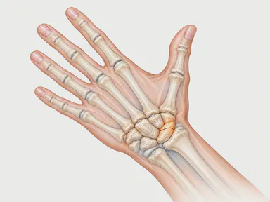
- Wrist ligaments Sprains and ruptures of the ligaments joining the carpal bones, especially the scapholunate ligament, which can destabilise the wrist and lead to osteoarthritis.
- Ganglion cyst A soft, benign, fluid-filled lump that arises from a joint or a tendon sheath, most often on the back of the wrist.
- Distal radius fracture The most common wrist fracture: it occurs on falling onto an outstretched hand and can be treated with a cast or with surgery depending on the displacement.
- Scaphoid fracture A fracture of the most vulnerable bone in the wrist: often not seen on the first X-ray, and if it fails to heal, it can lead to nonunion.
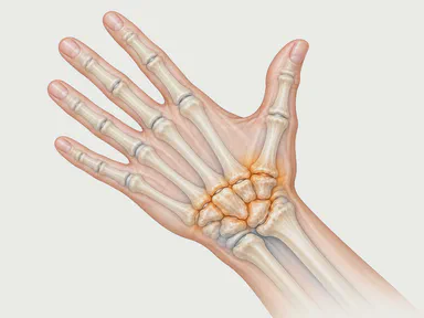
- Wrist osteoarthritis Wear of the wrist cartilage, almost always a consequence of an old fracture or ligament rupture, causing progressive pain and stiffness.
Sources and references
The information on this page is general and educational. It does not replace an individual medical assessment.
