

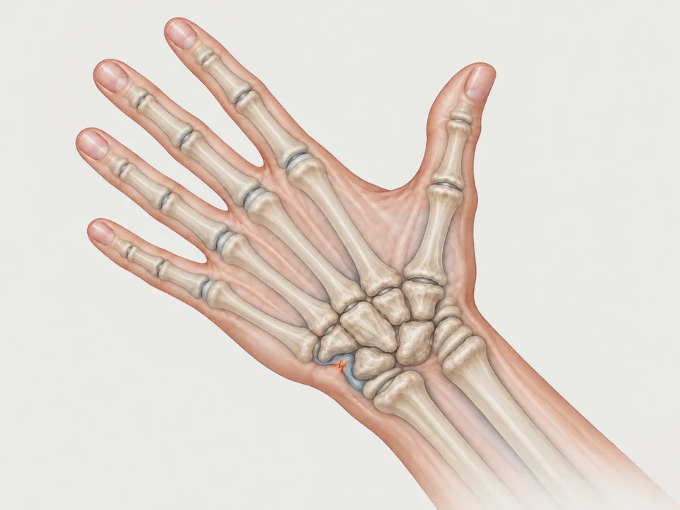
What is a triangular fibrocartilage injury?
The triangular fibrocartilage complex, known by its abbreviation TFCC, is a structure of cartilage and ligaments on the ulnar side of the wrist, that is, on the little-finger side. It extends between the edge of the radius and the head of the ulna and serves two functions: it cushions the loads passing from the carpus to the ulna, and it keeps the distal radioulnar joint, which allows the forearm to rotate, stable.
When the TFCC tears or wears down, pain appears on the ulnar side of the wrist and, in some cases, a sensation of instability when turning the hand. It is one of the most common causes of ulnar-sided wrist pain and is often diagnosed late, because X-rays are normal.
Symptoms
- Pain on the ulnar side of the wrist, in the gap between the ulna and the carpus, which increases when rotating the forearm, pushing up from a chair, or wringing out a cloth.
- Clicking or crunching when rotating the wrist.
- Loss of grip strength due to pain.
- In peripheral tears, a sensation that the head of the ulna is “popping out” or moving more than normal: this is distal radioulnar instability.
- Mild swelling in the ulnar area.
Causes and risk factors
Two types of injury are distinguished:
- Traumatic: caused by a fall onto the hand with the wrist in extension and rotation, by a sudden twist under load — for example with a drill that jams — or by a forced pull. These frequently accompany a distal radius fracture.
- Degenerative: caused by progressive wear, more common from middle age onwards and in people whose ulna is somewhat longer than the radius, which overloads the fibrocartilage with each weight-bearing movement. This is known as ulnar impaction syndrome.
Sports with repeated forearm rotation and load on the wrist, such as tennis, golf, gymnastics or rowing, favour both types.
Diagnosis
Clinical examination is fundamental. Pain on pressure over the ulnar fovea, rotation manoeuvres under load, and assessment of the stability of the distal radioulnar joint, compared with the healthy wrist, give strong guidance towards the diagnosis.
X-rays are used to rule out fractures and to measure the relative length of the ulna compared with the radius. MRI, and with greater precision MR arthrography with intra-articular contrast, shows most tears. Even so, the most reliable test is wrist arthroscopy, which allows direct visualisation of the fibrocartilage, assessment of its tension, and treatment during the same procedure.
Treatment
In recent injuries without instability, and in degenerative injuries, initial treatment is conservative:
- a wrist or forearm splint limiting rotation for a few weeks;
- adapting activities that cause pain;
- anti-inflammatories for short periods, if indicated;
- corticosteroid injection into the joint in selected cases;
- rehabilitation focused on the stability and strength of the wrist.
A significant proportion of people improve with these measures within two or three months.
When is surgery necessary?
Surgery is considered when:
- pain persists after several months of correct conservative treatment;
- there is instability of the distal radioulnar joint;
- the injury is traumatic, peripheral and recent in an active person, in whom repair offers a better chance of restoring stability;
- there is ulnar impaction with a long ulna that does not respond to treatment.
TFCC injury surgery
This is almost always performed by arthroscopy, through two or three incisions of only a few millimetres, as part of minimally invasive surgery of the wrist. The technique depends on the area affected:
- Tears in the central zone, which has almost no blood supply, do not heal even if sutured. In these, a debridement is performed: the torn edge is smoothed so that it stops catching and causing pain.
- Tears in the peripheral zone, next to the insertion on the ulna, do have a blood supply and can be repaired with sutures that reattach the fibrocartilage to the bone or capsule. This repair is what restores radioulnar stability.
- In ulnar impaction with a long ulna, the ulna can be shortened by a few millimetres with an osteotomy fixed with a plate, to unload the fibrocartilage.
Recovery
After arthroscopic debridement, the wrist is protected for a few days and mobility recovers within a few weeks. After a repair, the forearm is immobilised for around four to six weeks to protect the suture, followed by rehabilitation to regain rotation and strength. Return to load-bearing sport is usually delayed by several months. When the injury is degenerative or of long standing, the realistic goal is pain reduction, and the degree of improvement that can be expected is explained before surgery.
Specialist in TFCC injury in Barcelona
Dr Marta Martí Puente is a specialist in Orthopaedic Surgery and Traumatology with a focus on wrist surgery and arthroscopy. At her consultation in Barcelona she studies each case of ulnar-sided wrist pain individually, from conservative treatment through to arthroscopic debridement or repair of the triangular fibrocartilage, and supports recovery through to a return to activity.
Frequently asked questions
What does TFCC mean?
Does a TFCC injury heal on its own?
Does an MRI always detect the injury?
How long does recovery take after TFCC arthroscopy?
Related conditions
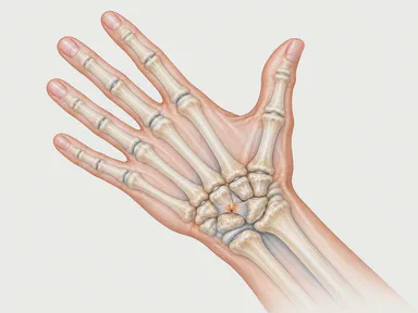
- Wrist ligaments Sprains and ruptures of the ligaments joining the carpal bones, especially the scapholunate ligament, which can destabilise the wrist and lead to osteoarthritis.
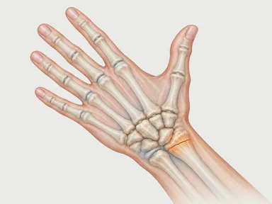
- Distal radius fracture The most common wrist fracture: it occurs on falling onto an outstretched hand and can be treated with a cast or with surgery depending on the displacement.
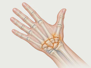
- Wrist osteoarthritis Wear of the wrist cartilage, almost always a consequence of an old fracture or ligament rupture, causing progressive pain and stiffness.
Sources and references
The information on this page is general and educational. It does not replace an individual medical assessment.
